Ob Medizin, Zahnmedizin oder Pharmazie: Organisiere Vorlesungen, PDFs und eigene Notizen nach Fach und erstelle daraus eine übersichtliche Zusammenfassung, die du bis zur Prüfung weiter verfeinerst.
Build one study note per subject.
Gather each subject’s key material into one structured note.
Focus on just the concept you need.
Open one concept, disease, or mechanism without searching the whole note.
Respiratory system notes
Respiratory system notesPulmonary function testsObstructive ventilatory defect
Respiratory system notes
Review respiratory physiology and pulmonary function tests in one note.
Connect obstructive patterns with their key spirometry findings.
Alveolar ventilation
Calculate effective ventilation after dead space and connect it with PaCO₂.
Gas exchange
Separate diffusion limitation, V/Q mismatch, and shunt when evaluating hypoxemia.
Pulmonary function tests
Use FEV₁/FVC and bronchodilator response to organize obstructive patterns.
Obstructive ventilatory defect
Increased airway resistance lowers FEV₁/FVC and prolongs expiration.
A reduced FEV₁/FVC ratio is the key finding.
Expiration is prolonged and air trapping can develop.
Interpret bronchodilator response together with symptom change.
Spirometry
Confirm both a reduced FEV₁ and a low FEV₁/FVC ratio in an obstructive pattern.
AsthmaReversible airway obstructionWorse at night or after exercise
COPDPersistent airflow limitationSmoking history and chronic cough
Obstructive ventilatory defect
Increased airway resistance lowers FEV₁/FVC and prolongs expiration.
A reduced FEV₁/FVC ratio is the key finding.
Expiration is prolonged and air trapping can develop.
Interpret bronchodilator response together with symptom change.
Spirometry
Confirm both a reduced FEV₁ and a low FEV₁/FVC ratio in an obstructive pattern.
AsthmaReversible airway obstructionWorse at night or after exercise
COPDPersistent airflow limitationSmoking history and chronic cough
See the whole note as a mind map.
Zoom out to see every section and how the subject fits together.
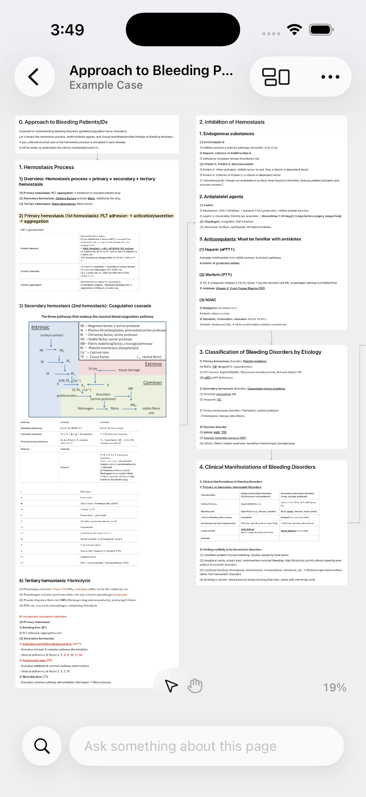
Bleeding disorders review
Link hemostasis, screening tests, and differential clues
Read the bleeding pattern before the laboratory pattern
Before exams, scan pathways, test patterns, and next steps
Primary hemostasis
Vessel injury → vasoconstriction → platelet plug
vWF–GPIb mediates platelet adhesion
ADP and TXA₂ amplify platelet activation
GPIIb/IIIa–fibrinogen drives aggregation
Mucosal bleeding and petechiae suggest this pathway
Secondary hemostasis
Coagulation factors generate thrombin and fibrin
Intrinsic: XII → XI → IX + VIII
Extrinsic: tissue factor + VII
Common: X + V → II → I
Fibrin stabilizes the initial platelet plug
Initial hemostasis tests
CBC/platelets · PT/INR · aPTT · fibrinogen
PT screens extrinsic and common pathways
aPTT screens intrinsic and common pathways
Bleeding pattern → medication → test pattern
Normal screens do not exclude every mild disorder
Coagulation cascade
Intrinsic and extrinsic arms converge at factor X
Xa + Va convert prothrombin to thrombin
Thrombin converts fibrinogen to fibrin
Factor XIII cross-links and stabilizes fibrin
PT/aPTT pattern
Use the prolonged test to localize the pathway
PT only: factor VII or extrinsic-pathway clue
aPTT only: VIII · IX · XI · XII or inhibitor
Both: common pathway · liver · DIC · medication
Interpret platelet count and fibrinogen together
Mixing study
Mix patient plasma with normal plasma 1:1
Correction suggests a clotting-factor deficiency
No correction suggests an inhibitor or medication
Incubation can reveal a time-dependent inhibitor
Confirm with factor and inhibitor assays
Factor deficiency
Prolonged aPTT corrects after mixing
Measure factors VIII · IX · XI as indicated
Hemophilia A/B are key inherited examples
Connect results with personal and family history
Inhibitor present
Prolonged aPTT fails to correct
Consider lupus anticoagulant or a factor inhibitor
Exclude anticoagulant medication interference
Use confirmatory assays with the clinical context
Spread out confusing concepts on the canvas.
When notes make the overall flow hard to follow, spread concepts out to see connections.
Respiratory system notes
Ask AI whenever something is unclear.
Get answers grounded in your uploaded PDFs, then add them to your note.
Respiratory system notesObstructive ventilatory defect
Obstructive ventilatory defect
Higher airway resistance lowers FEV₁/FVC and slows expiration.
Spirometry
Check for a full inhale, forceful exhale, and repeatable curves. FVC is total exhaled volume; FEV₁ is the volume exhaled in one second. Confirm test quality before interpreting the values.
FEV₁/FVC interpretation
Obstruction lowers both FEV₁ and the FEV₁/FVC ratio. A ratio below the age-adjusted lower limit supports airflow obstruction.
Compare FEV₁ with the expiratory curve. A concave curve and lower peak flow support obstruction.
If FVC is also low, confirm restriction with lung volumes. Compare severity with symptoms and earlier results.
Bronchodilator response
Compare FEV₁ and FVC before and after a bronchodilator. A clear FEV₁ recovery suggests reversible airway obstruction.Add to chat
A positive response supports variable airflow, but does not diagnose asthma alone. If negative, repeat the test when symptoms are present. Interpret it with symptom changes and repeat measurements.
Review your notes wherever you study.
Organize on desktop, then use the upcoming mobile app for quick reviews anywhere.
Turn notes into quizzes, PDFs, and Word files.
Make a quiz, export cards to Anki, or save your notes as PDF or Word.
Respiratory system notes
Alveolar ventilation
Alveolar ventilation accounts for dead space when relating tidal volume to respiratory rate. Rapid, shallow breathing can reduce gas exchange even when minute ventilation appears unchanged.
VA = (VT - VD) × RR
Hypoventilation can raise PaCO₂ and cause respiratory acidosis.
Gas exchange and the oxygen dissociation curve
O₂ diffuses from alveoli to blood, while CO₂ moves from blood to alveoli. Lower pH, higher CO₂, and higher temperature shift the oxygen dissociation curve to the right.
Right shift: oxygen is released to tissues more easily.
Left shift: hemoglobin holds oxygen more tightly.
Causes of hypoxemia
Compare V/Q mismatch, diffusion defect, shunt, and hypoventilation using the A–a gradient and response to supplemental oxygen.
V/Q mismatch: increased A–a gradient and usually improves with oxygen.
Shunt: PaO₂ improves only slightly despite oxygen.
Hypoventilation: PaCO₂ rises while the A–a gradient may stay normal.
Obstructive ventilatory defect
Asthma and COPD characteristically reduce FEV₁/FVC. Expiration becomes prolonged, air trapping develops, and residual volume can increase.
Chronic bronchitis: productive cough for at least 3 months in 2 consecutive years.